29+ Cystic Hygroma In Fetus Pictures
Cystic Hygroma In Fetus Pictures. The cause is unknown but may be related to genetic changes in the fetus. If your baby has normal chromosomes and the cystic hygroma disappears by 20 weeks of pregnancy, the outcome will probably be good.

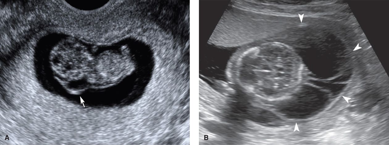
All the women denied previous pregnancies with cystic hygroma. The fetus shown here died in utero (intrauterine fetal demise) and shows signs of maceration (autolysis) such as the slippage of the skin and the reddened color. Doctors usually diagnose cystic hygromas when the fetus is still in the womb, often during a routine abdominal ultrasound.
eau de toilette spray for men disque meuleuse pour bois dalle adhesive leroy merlin dessin couronne de noel a colorier
Fetal Imaging Obgyn Key
The lymphatic system is a network of vessels that maintains fluids in the blood, as well as transports fats and immune system cells. Instead, your doctor will closely monitor your baby’s health. Cystic hygroma, also known as cystic or nuchal lymphangioma, refers to the congenital macrocystic lymphatic malformations that most commonly occur in the cervicofacial regions, particularly at the posterior cervical triangle in infants. If your baby has normal chromosomes and the cystic hygroma disappears by 20 weeks of pregnancy, the outcome will probably be good.

When you’re pregnant, your doctor may find your baby’s cystic hygroma during a routine ultrasound.these cysts are. It is a congenital defect that can affect any part of the human body but it in most cases affects the neck and head. This leads to the formation of cysts, pockets of fluid beneath the baby’s skin, like big blisters. Will the.

They will indicate whether an abnormality exists. Gallagher et al., 1999).this finding is to be differentiated from simple increased nuchal translucency in which skin thickening is noted at the posterior aspect of the fetal neck. Such material is called embryonic lymphatic tissue. A communication between the primitive lymphytic system and the jugular vein is formed in the early gestation. Cystic.

A baby with no other health problem and a small cystic hygroma will be observed by ultrasound every three to four weeks. As the baby grows in the womb, it can develop from pieces of material that carries fluid and white blood cells. If your baby has normal chromosomes and the cystic hygroma disappears by 20 weeks of pregnancy, the.

View 61 kb version one very characteristic feature of a fetus with monosomy x is the cystic hygroma of the neck. Health concerns for both the developing baby and the pregnant woman. A communication between the primitive lymphytic system and the jugular vein is formed in the early gestation. When a cystic hygroma goes away, the developing baby’s chance for.

All the women denied previous pregnancies with cystic hygroma. A baby with no other health problem and a small cystic hygroma will be observed by ultrasound every three to four weeks. Most of the cystic hygromas are noticeable as the child reaches 2 years old. Most fetuses with cystic hygroma have abnormal karyotype. As the baby grows in the womb,.

The ultrasound machine used here is the toshiba aplio 500 system. Will the cystic hygroma go away? This ultrasound imaging study of fetal multiple cystic hygromas of the fetus are courtesy of dr. A cystic hygroma is a type of birth defect. It can be visible as the child grows older and the ch can also get larger and bigger.

Cystic hygroma refers to the abnormal lymphatic lesion that mostly develops at birth. Cystic hygroma, also known as cystic or nuchal lymphangioma, refers to the congenital macrocystic lymphatic malformations that most commonly occur in the cervicofacial regions, particularly at the posterior cervical triangle in infants. A cystic hygroma is also known as a lymphatic malformation. A cystic hygroma is a.

A cystic hygroma is the result of toxins being unable to leave the baby’s body. Symptoms and signs include the appearance of. The mean size of the fetal cystic hygroma at diagnosis was 7.9 mm (range: Doctors may also detect it. As the baby grows in the womb, it can develop from pieces of material that carries fluid and white.